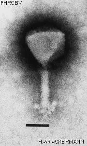
HER 165
Add to cart
Name
VP1
Morphotype
A1 (Myophage)
Realm
Duplodnaviria
Kingdom
Heunggongvirae
Phylum
Uroviricota
Class
Caudoviricetes
Electron Micrograph
Image

Image description
Magnification: 297,000X
Bar: 50 nm
Staining: PTB
Bar: 50 nm
Staining: PTB
Characteristics
Plaques : 0.2-0.3 mm, clear.
Collar fibers.
Plaques easier to see after 3 days.
Genomic sequence
No
Bacterial hosts
HER
Reference
Koga, T., S. Toyoshima, and T. Kawata. 1982. Morphological varieties and host ranges of *Vibrio parahaemolyticus* bacteriophages isolated from seawater. Appl. Environ. Microbiol. 44:466-470.
History
Received from
Date
Dr Tomio Kawata,
Department of Food Microbiology,
Tokushima University School of Medecine,
Tokushima,
770,
Japan.
Department of Food Microbiology,
Tokushima University School of Medecine,
Tokushima,
770,
Japan.
02-21-1983
Isolated by
Date
T. Koga and T. Kawata
Tokushima, Japan
Tokushima, Japan
08-1977
Source
sea water
Updated at
2024-01-16