HER 202
Add to cart
Name
I3
Morphotype
A1 (Myophage)
Realm
Duplodnaviria
Kingdom
Heunggongvirae
Phylum
Uroviricota
Class
Caudoviricetes
Genus
Bixzunavirus
Species
Bixzunavirus I3
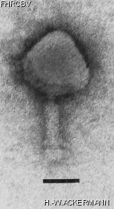
Electron Micrograph
Image

Image description
Magnification: 297,000X
Bar: 50 nm
Staining: UA
Bar: 50 nm
Staining: UA
Characteristics
Temperate and transducing phage.
Turbid plaques.
The only Mycobacterium phage with a contractile tail.
Life cycle of 5 hours.
Genomic sequence
Yes
Bacterial hosts
HER
Reference
Kozloff, L.M., C.V.S. Raj, R.N. Rao, V.A. Chapman, and S. Delong. 1972. Structure of a transducing mycobacteriophage. J. Virol. 9:390-393.
Remarks
Propagate the phage at 33°C.
History
Received from
Date
Dr. K.P. Gopinathan
Microbiology and Cell Biology Laboratory
Indian Institute of Science
Bangalore 560012, India
Microbiology and Cell Biology Laboratory
Indian Institute of Science
Bangalore 560012, India
12-12-1983
Isolated by
Date
C.V. Sundar and T. Ramakrishnan
Microbiology and Cell Biology Laboratory
Indian Institute of Science
Microbiology and Cell Biology Laboratory
Indian Institute of Science
1969
Source
Soil, Bangalore, India
Updated at
2024-01-16