HER 86
Add to cart
Name
1A
Morphotype
B1 (Siphophage)
Realm
Duplodnaviria
Kingdom
Heunggongvirae
Phylum
Uroviricota
Class
Caudoviricetes
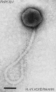
Electron Micrograph
Image

Image description
Magnification: 297,000X
Bar: 50 nm
Staining: UAB
Bar: 50 nm
Staining: UAB
Characteristics
Plaques: 0.5mm, clear.
Collar present or not.
Morphogically identical with phage 2A (2070).
Part of typing set.
Genomic sequence
No
Bacterial hosts
HER
Reference
Yousten, A. A., H. de Barjac, J. Hedrick, V. Cosmao Dumanoir, and P. Myers. 1980. Comparison between bacteriophage typing and serotyping for the differentiation of *Bacillus sphaericus* strains. Ann. Nicrobiol. 131B: 297-308.
Remarks
Propagation at 32°C.
Set of phage measure taked in ME are included in the original form.
History
Received from
Date
Dr A. Yousten,
Biology Department,
Virginia Polytechnic Institute and State University, Blacksburg, Va 24061,
USA.
Biology Department,
Virginia Polytechnic Institute and State University, Blacksburg, Va 24061,
USA.
28-04-1982
Isolated by
Date
Dr J. Hedrick,
Biology Department (now Food Science),
Blackburg, VA,
USA.
Biology Department (now Food Science),
Blackburg, VA,
USA.
1978
Source
Soil, Blackburg, VA, USA.
Updated at
2024-01-16