HER 97
Add to cart
Name
PBS1
Morphotype
A1 (Myophage)
Realm
Duplodnaviria
Kingdom
Heunggongvirae
Phylum
Uroviricota
Class
Caudoviricetes
Genus
Takahashivirus
Species
Takahashivirus PBS1
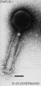
Electron Micrograph
Image

Image description
Magnification: 297,000X
Bar: 50 nm
Staining: UAB
Bar: 50 nm
Staining: UAB
Characteristics
Plaques: 0.1, clear to veiled.
Helical and contraction fibers, very large size
Thymine replaced by uracil.
Generalized transduction
Very stable
Takahashi: Penassay broth
Tryptose blood agar base
Schaefferʼs spore medium.
Genomic sequence
Yes
Bacterial hosts
HER
Reference
Eiserling, F. A. 1963. *Bacillus subtilis* bacteriophages: structure, intracellular development and conditions of lysogeny. Ph.D. thesis, University of California, Los Angeles. pp. 1-127.
Remarks
Description of the media used are available upon request.
History
Isolated by
Date
Dr. I. Takahashi,
Departement of Biology,
McMaster University
Departement of Biology,
McMaster University
1960
Received from
Date
Dr. I. Takahashi,
Departement of Biology,
McMaster University,
Hamilton, ON.
Canada, L8S 4K1
Departement of Biology,
McMaster University,
Hamilton, ON.
Canada, L8S 4K1
5-04-1982
Source
Soil, Ottawa, Canada
Updated at
2024-01-16