HER 393
Add to cart
Name
P1D
Morphotype
A1 (Myophage)
Family
?1
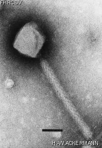
Electron Micrograph
Image

Image description
Magnification: 297,000X
Bar: 50 nm
Staining: PTB
Bar: 50 nm
Staining: PTB
Characteristics
Plaques are imperceptible: 0.1 mm, veiled.
Genomic sequence
No
Bacterial hosts
HER
Reference
Ackermann, H.-W. and coll. 1994. P1D, a new member of the P1 bacteriophage group.
Remarks
Variant of P1; Slightly longer tail, almost no head size variants, different host range and plaque size.
Plaques easier to observe at 42°C than at 37°C; also produced spontaneously.
Other host strains: HER 1128(*E. coli* O15), Cubizolles, blanchon, LY265.
Plaques easier to observe at 42°C than at 37°C; also produced spontaneously.
Other host strains: HER 1128(*E. coli* O15), Cubizolles, blanchon, LY265.
History
Isolated by
Date
A. Reynaud and H.-W. Ackermann
Laboratory of Ackermann
Laboratory of Ackermann
12-1991
Source
*E. coli* O103 4472 (also strains GVs and 2198) after UV + mitomycin C induction.
Updated at
2024-01-22